- Home
-
Products
-
 Antibodies
- CD Antibodies
- IL Antibodies
- Active Antibodies
- Point Mutation Antibodies
- AD Related Antibodies
- Tag Antibodies
- Primary Antibodies
- Secondary Antibodies
- G Protein Monoclonal Antibodies
- G Protein Polyclonal Antibodies
- Loading Control Antibodies
- Biosimilar Antibodies
- Flow Cytometry Antibodies
- Monoclonal Antibodies
- Polyclonal Antibodies
-
 Kits
Kits
-
 Peptides
-
 Proteins
-
Biochemicals
-
- Services
- Featured Products
- Research Area
- FAQ
- News
- About Us
- Contact Us
CD Antibodies
Products
Products
Mouse Monoclonal Antibody to CD42B
All
- Catalog: TD-31457
- Clonality: mAb
- Application: WB, FCM
- Synonyms: GP1BA; BSS; GP1B; VWDP; GPIbA; BDPLT1; BDPLT3; DBPLT3; GPIbalpha; CD42b-alpha
-
Specification:
100ul 50ul
-
Prices:
¥2180
Mouse Monoclonal Antibody to CD42B
|
|
| Description | |
| Glycoprotein Ib (GP Ib) is a platelet surface membrane glycoprotein composed of a heterodimer, an alpha chain and a beta chain, that is linked by disulfide bonds. The Gp Ib functions as a receptor for von Willebrand factor (VWF). The complete receptor complex includes noncovalent association of the alpha and beta subunits with platelet glycoprotein IX and platelet glycoprotein V. The binding of the GP Ib-IX-V complex to VWF facilitates initial platelet adhesion to vascular subendothelium after vascular injury, and also initiates signaling events within the platelet that lead to enhanced platelet activation, thrombosis, and hemostasis. This gene encodes the alpha subunit. Mutations in this gene result in Bernard-Soulier syndromes and platelet-type von Willebrand disease. The coding region of this gene is known to contain a polymophic variable number tandem repeat (VNTR) domain that is associated with susceptibility to nonarteritic anterior ischemic optic neuropathy. | |
| Specification | |
| Aliases | GP1BA; BSS; GP1B; VWDP; GPIbA; BDPLT1; BDPLT3; DBPLT3; GPIbalpha; CD42b-alpha |
| Entrez GeneID | 2811 |
| Swissprot | P07359 |
| clone | 6H6H10 |
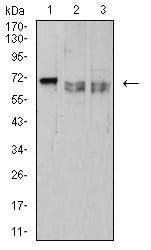
| WB Predicted band size | 71.5kDa |
| Host/Isotype | Mouse IgG1 |
| Antibody Type | Primary antibody |
| Storage | Store at 4°C short term. Aliquot and store at -20°C long term. Avoid freeze/thaw cycles. |
| Species Reactivity | Human |
| Immunogen | Purified recombinant fragment of human CD42B (AA: extra 17-183) expressed in E. Coli. |
| Formulation | Purified antibody in PBS with 0.05% sodium azide |
| Application | |
| WB | 1/500 - 1/2000 |
| IHC | 1/200 - 1/1000 |
| FCM | 1/200 - 1/400 |
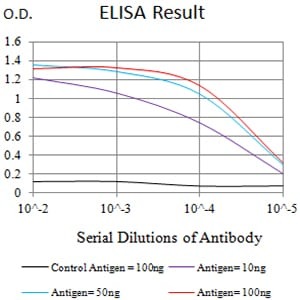
| ELISA | 1/10000 |
| Product Image | |
|
|
|
|
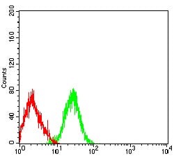
| Black line: Control Antigen (100 ng);Purple line: Antigen (10ng); Blue line: Antigen (50 ng); Red line:Antigen (100 ng) | Western blot analysis using CD42B mouse mAb against HL-60 (1), MOLT4 (2), and Ramos (3) cell lysate. | Flow cytometric analysis of HL-60 cells using CD42B mouse mAb (green) and negative control (red). |
|
|
||
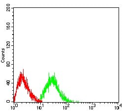
| Flow cytometric analysis of Jurkat cells using CD42B mouse mAb (green) and negative control (red). |